Sasha and I
About
I'm Jasmin and I love dogs. I was born and raised in Jerusalem, where I spent my time either in school, Schmidt's Girls' College, at the beach in Herzliya, or playing tennis. I left to the United States when I was 16, where I attended the United World College in New Mexico (UWC-USA) and had the time of my life. New Mexico is unlike any other place I have ever seen. I then went to the University of Pennsylvania, where I studied chemical engineering, fell in love with Parc's bread, and experienced the painful joy of an 'all-nighter'. In my last year at Penn, I worked with Prof. Gorte and his graduate student at the time, Kevin Bakhmutsky. They made me fall in love with research and detective work and I was under great mentorship. So I applied for graduate school, and I've been at Princeton since 2012, still under great mentorship in the Shvartsman Lab.
Research
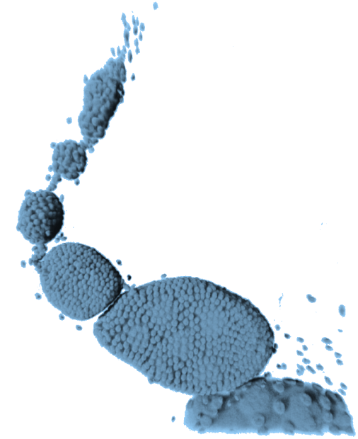
I am currently working on the egg chamber in female fruit flies, which is a relatively simple multicellular structure, and a precursor to the oocyte or mature egg. It's a great system for understanding a lot of basic biological phenomena at the single-cell level, as well as collective and emergent behaviors that arise in small multicellular systems. Egg chambers grow in these assembly lines, like the one shown below, and remain connected throughout their entire developmental period in the fly's abdomen. So by dissecting out one of these assembly lines, we get a snapshot of all the different stages of development a single egg chamber is going to go through. The nuclei of each cell are shown in red below.
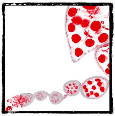
I am looking into the dynamics of collective growth on a small lineage tree, and the diversity of its 3D packing configurations. We also just started a new project looking at how growth is coordinated and regulated in a system with two different cell types.
-
On Growth
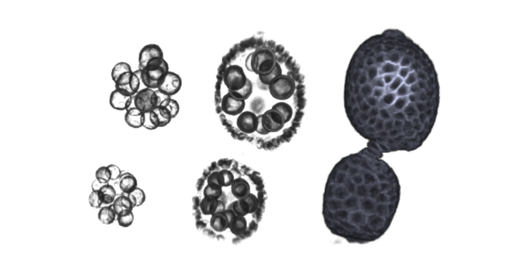
In Drosophila, oocytes develop within a multicellular structure comprising 16-cell germ cells and an overlying layer of epithelial cells. The germline cells, 1 oocyte and 15 nurse cells, are stereotypically connected by membranous bridges that allow intercellular communication and transport. These are called ring canals and are shown below as red rings. Throughout oogenesis, the nurse cells experience a dramatic increase in size, become polypoid, and produce whatever the oocyte and future embryo require for growth. Their nuclei become relatively huge: these are the large gray circles in the same image below. One of the most interesting observations of nurse cell growth has remained unquantified and unexplained: the initial seemingly uniform distribution of cell sizes becomes increasingly polarized as oogenesis proceeds, with the cells most adjacent to the oocyte nearly double the size of those furthest away. Studies of mutants suggest a connection between distance from the oocyte and nurse cell growth, however, confounding factors exist.
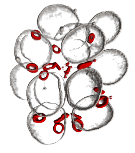
We therefore asked: can we quantify this pattern of cell sizes, and can a simple model of interconnected cells that allows for growth and exchange of cellular components, explain the emergent pattern of cell sizes in developing egg chambers? This work was recently presented at an EMBO workshop on cell size regulation, and became the subject of our recent Current Biology paper, "Collective Growth in a Small Cell Network".
-
On Form
Packing problems are pervasive, from the stacking of oranges at the grocer's and Guinness casks on a ship, to the formation of honeycombs, and self assembly of viral protein coats. Since Kepler, people have been interested in coming up with principles that account for the observed packing configurations of infinite and finite collections of spherical and non-spherical objects.
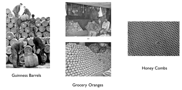
Similarly, we're interested in describing and understanding the packing configurations of the 16 cells comprising the Drosophila egg chamber, and in coming up with 'packing principles' that explain our observations. Using both experimental and computational approaches, we enumerated and characterized the complete set of permissible packing configurations, and their relative frequency. To do so, we relied on z-stacks of labelled egg chambers, and uniquely mapped their 3D structure onto the various configurations of the ring canal tree. In doing so, we reduced high dimensional data to a 1D descriptor that captures most of the cells' spatial organization: the configuration of the lineage tree, depicted as a collection of nodes and edges, uniquely captured the egg chambers three dimensional configuration. This study emphasizes the indispensability of insightful visualization and abstraction techniques to pattern extraction. This work then took a more theoretical turn and is currently in review at Nature Physics.
This idea of using low dimensional representation of multidimensional data to extract patterns was featured in a Nature news and views showcasing transformative techniques and toolboxes for visualizing biology.

I also had the opportunity to present at a conference / workshop on visualization biological data (VIZBI) in Heidelberg this year, where participants were invited to submit an artistically inspired picture related to their work. Stas, my advisor, had earlier sent me an image of Picasso's series of lithographies, titled Bull, depicting .... a bull at various stages of abstraction. The first image was of a 'meaty' bull, and the last, was just a collection of lines that were still unmistakably a bull - an abstraction of the bull. The analogy with our work was unavoidable: despite reducing the 3D image of an egg chamber to collection of nodes and edges, the resulting tree captured the essence of that egg chamber's spatial organization, i.e. the 'spirit of the beast'. I therefore created this montage showing a few of Picasso's sequential reductionist lithographs, and our own series of images. The idea was not draw direct comparisons, but to use well known pieces of art to make it easier to convey our approach and our message to those unfamiliar with our work, and I think it worked. People liked it, and the image was awarded the Autodesk Art and Biology Award.

All of the above work was done in collaboration with Paul Villoutreix, a previous postdoc at our lab, Alexander M.Berezhkovskii from NIH, Norbert Stoop from the Dunkel lab at the Math department at MIT. Unwavering gratitude to my advisor, Stas Shvartsman and members of my committee, Yannis Kevrekidis, Celeste Nelson, and Clifford Brangwynne from CBE.
-
Hindgut Organoids
Organoids are cell aggregates that arise in vitro with 3D structure, which more closely mimics the in vivo tissue than 2D models. Given the proper environment and growth factors, pluripotent stem cells will differentiate into organoids of several tissues including brain, liver and intestine. A mechanistic understanding of early organoid formation is essential for transitioning these technologies into robust, high-throughput methods suitable for commercial and therapeutic applications. In the past two years, I've been involved in an EBICS project that aims to understand the mechanisms regulating the emergence of intestinal organoids from hiPSCs derived hindgut cultures, and to design systems that sort for organoids predicted to mature successfully. This work is central to translating this technology into high-throughput models for drug discovery and development.

A hindgut organoid stained to reveal nuclei and different cell type (epithelial and mesenchymal)
This work was done in collaboration with the Kamm (MIT), Griffith (MIT) and Asada (MIT) labs, specifically with Natasha Arora and Jacob Guggenheim. A manuscript reporting these studies, titled “A process engineering approach to increase organoid yield” in Development can be found here.
Art & Communicating Science
In addition to being a great model system, the egg chamber is also a beautiful structure. Most of my work requires 3D images, or z-stacks, or egg chambers with multiples structures labeled simultaneously. The images and the colors are fantastic and make it easy to get others excited about one's work, and about science in general.
I realized this very early on and got involved in the Art of Science exhibit, held (almost) annually here at Princeton, initially as a participant, now as an organizer. It's an exhibit showcasing some of the unintentionally beautiful images and videos captured in the process of doing science, and is a great opportunity to engage the public in research seemingly going on behind closed doors.
Please visit our 2017 exhibit at the Friend Center Atrium
Weekdays, 9 am - 5 pm. On view through April 2018
I also come from an artistically inspired family. My mother, Ursula, and younger sister, Besan, have a taken on a mammoth project of putting together an art exhibit of their works. It is still a work in progress, but if you're interested, you can visit their website that hosts pictures of their pieces, or even contact them !
Outreach & Writing
I love doing science and telling others about it. I've recently become involved with The Node as an author, which is just a great platform for all things developmental biology. There are lists of events, conferences, and more importantly, relatively informal contributions from scientists about their work. Most interestingly are the blogs that highlight aspects of research that did not make it into the paper but were germaine to the work itself. Here's the piece I wrote about our work on collective growth.
I also wrote about growth, more informally, on Massive. I love reading about growth and what regulates this incredibly intuitive, yet amazingly complex phenomenon. If you have questions about growth or think I should write about a particular aspect of it that interests you, please let me know !
Do you know why you stop growing for example? Why are some dogs huge, others tiny?

Huge Sasha, Tiny Zeus
Last but not least, I recently had the opportunity to participate in a great outreach program called Skype a Scientist. Truely, if you have 30 minutes to spare, do participate: you get to tell school kids of all ages from anywhere in the world about what you do, and hopefully get them excited about it. We need advocates for science. Here's what I wrote about my experience.
Education at a Glance
- 2012-Present | Princeton University
PhD Candidate in Chemical & Biological Engineering
- 2007-12 | University of Pennsylvania
MSE & BSE in Chemical & Biomolecular Engineering
Minor in Mathematics and Materials Science & Engineering
Concentration in Environmental Engineering
- 2005-07 | United World College (UWC-USA, NM)
International Baccalaureate (IB), hot springs and the worldliest of places
- 1993-05 | Schmidt’s Girls’ College, Jerusalem
Typing lessons. Mathematics in Arabic and in English.
Please contact me for a complete resume or with any questions in general :)
.... and this is my dog Zeus
